
Immunohistochemistry in Cancer Diagnosis
By Raymund Bueno, PhD
Immunohistochemistry (IHC) is a method performed in the lab to identify specific antigens or analytes in a tissue sample using monoclonal or polyclonal antibodies¹ (see Figure 1). This technique was first used in 1941 by Coons et al.² When using immunohistochemistry in cancer diagnosis, there are specific antigens that are unique or over-expressed. Immunohistochemistry is used to look at these antigens, also known as analytes, to answer a number of research and clinical questions including (a) distinguish a tissue as being cancerous, (b) assess whether a patient should receive or is responding to therapy, or (c) evaluate the immune response at the site of a tumor. To perform immunohistochemistry, a tissue biopsy is formalin-fixed and paraffin embedded before being processed into sections and fixed onto slide. The slides are incubated with an antibody that will bind to the antigen of interest. The sites of antibody and antigen binding in the tissue are visualized using fluorescent or light microscopes, depending on the type of antibody used.
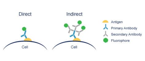
Figure 1
An improved version of immunohistochemistry is multiplexed immunohistochemistry. This technique detects more than one antigen on a single slide, allowing for a deeper and more complex visual of a tissue. Multiplex IHC can provide insights into tumor heterogeneity and the tumor microenvironment.³ However, multiplex IHC can be difficult to interpret as the fluorescence from the different antibodies or non-specific binding can lead to complications in visualizing the results. Further, there are limitations on the number of antibodies/fluorophores that may be used, so this technique is still not considered a high-throughput analysis. Finally, although advancements in image analysis software has delivered improved quantitation, most IHC techniques are still considered only semi-quantitative, at best.
Cofactor’s ImmunoPrism assay was designed to overcome some of the challenges of immunohistochemistry in cancer diagnosis. ImmunoPrism is built on Predictive Immune Modeling, which uses multidimensional modeling of RNA to better understand and diagnose cancer. Unlike IHC or multiplex IHC, ImmunoPrism leverages high-throughput analysis of gene expression, rather than relying on antibody binding and microscopy. This allows for deeper and more quantitative molecular insights into the disease by comparing the RNA expression of the tissue to Cofactor’s databae of Health Expression Models (currently focused on immune cells). Similar to multiplex IHC, the ImmunoPrism assay takes a multi-analyte approach to gain a deeper realization into the complexity of cancer. Analyzing solid tumor materials both with the ImmunoPrism assay and multiplex IHC can provide researchers and clinicians with the best of both worlds – high-through and quantitative assessment through ImmunoPrism, with spatial information provided with IHC. This combination enables a more accurate representation of biological signals and their interconnectedness, allowing for more impactful interpretations.
- Duraiyan J, Govindarajan R, Kaliyappan K, Palanisamy M. Applications of immunohistochemistry. J Pharm Bioall Sci 2012;4, Suppl S2:307-9
- Coons, AH, Creech, HJ, Jones, RN. Immunological properties of an antibody containing a fluorescent group. Proc Soc Exp Biol Med. 1941;47(2):200–202.
- Stack, E. C., Wang, C., Roman, K. A., & Hoyt, C. C. (2014). Multiplexed immunohistochemistry, imaging, and quantitation: a review, with an assessment of Tyramide signal amplification, multispectral imaging and multiplex analysis. Methods, 70(1), 46-58.

